
Project 3
Cynthia K. Thompson, Ph.D. and her research team investigate the effect of sentence-level treatment on language and brain recovery. Participants receive treatment focused on complex sentence structures, with recovery of untrained less complex sentences an expected outcome of treatment. Processing routines used as people comprehend and produce sentences are examined using eye-tracking techniques and the neural mechanisms of recovery are examined using neuroimaging methods.
Dr. Thompson is Ralph and Jean Sundin Professor of Communication Science in the Roxelyn and Richard Pepper Department of Communication Sciences and Disorders at Northwestern University. She trained in Psychology, Linguistics, and Speech andLanguage Pathology. She is world-renowned for her research on aphasia, brain plasticity, and language processing. She is the Director of the Center for the Neurobiology of Language Recovery, and an expert and leading authority on sentence processing disorders.
Northwestern University Aphasia and Neurolinguistics Research Website
Current Projects:
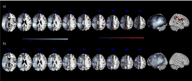
Neural plasticity of language and domain-general networks in aphasia. In this paper, we examined the effects of treatment on both offline and online sentence processing and associated neuroplasticity within language and domain-general networks in chronic stroke-induced agrammatic aphasia. Nineteen aphasic individuals were randomly assigned to either receive a 12-week course of linguistically-based treatment of passive sentence production and comprehension (Mack & Thompson, 2017) or to serve as control participants. Both aphasic groups performed two offline behavioral tasks at the start and again three months following (at post-testing). Participants also performed an online eyetracking comprehension task and a picture-verification fMRI task. Our results showed that individuals in the treatment, but not in the control (no treatment), group improved on production and comprehension of trained structures, and that treatment effects also generalized to untrained syntactically similar structures. At post-testing, participants in the treatment (and not in the control) group also showed eye movement patterns that were more similar (compared to pre-testing) to the patterns found in healthy individuals on the same task (Mack & Thompson, 2017). Treatment also resulted in activation increases in right hemisphere regions that encompassed both language and domain-general regions (Figure 1), and changes in activation were positively correlated with changes in both offline and online sentence comprehension. Overall, these findings indicate that – when the sentence processing network in the left hemisphere is disrupted – the right hemisphere can be recruited to compensate for language deficits.
Artificial Grammar Learning (AGL). Through this experiment, we (Schuchard & Thompson, 2017) examined AGL in older (44 – 74 yrs.) healthy adults (n=36; 24 randomly assigned to a trained group, with 12 untrained) and a group of agrammatic aphasic individuals (n=12) using an artificial grammar adapted from “Language P” (Saffran, 2002). Monosyllabic pseudowords, also known as one syllable fake words, were assigned to one of five lexical categories and were arranged according to the rules of a hierarchical phrase structure grammar to create pseudoword sentences that were used for auditory exposure-based training. Participants underwent a grammaticality judgment test at three time points: immediately following training (Test 1), one day after training (Test 2), and after a second training session (Test 3). Results showed significantly greater learning for both trained groups (healthy and aphasic) compared to the untrained healthy participant group and greater gains at Test 3 vs. Test 2 were found for healthy (than for aphasic) participants.
Eyetracking Reliability. Because we sought to use eyetracking to measure the effects of treatment in Cycle 1, we (Mack et al., 2016) first examined the stability of eye movements over time in 21 young controls and 12 adults with aphasia using a sentence-picture matching task (after Meyer et al., 2012). Participants performed the task on two separate occasions, one week apart. Both groups showed little eye movement variability across test points, with Intraclass correlations (ICCs) for the aphasic group in the good and excellent range for actives and passives (sentence end), respectively.

Meta-analysis of Neural Activation for Sentence Comprehension and Production (Neurotypical Adults). We examined three issues related to the brain networks underlying sentence comprehension and production in healthy individuals: First, which regions are recruited for sentence comprehension and sentence production? Second, are there differences for auditory sentence comprehension vs. visual sentence comprehension? Third, which regions are specifically recruited for the comprehension of syntactically complex sentences? Results from activation likelihood estimation (ALE) analyses (from 45 studies) implicated a sentence comprehension network occupying bilateral frontal and temporal lobe regions. Regions implicated in production (from 15 studies) overlapped with the set of regions associated with sentence comprehension in the left hemisphere, but did not include inferior frontal cortex, and did not extend to the right hemisphere. Modality differences between auditory and visual sentence comprehension were found principally in the temporal lobes. Results from the analysis of complex syntax (from 37 studies) showed engagement of left inferior frontal and posterior temporal regions, as well as the right insula. The involvement of the right hemisphere in the comprehension of these structures has potentially important implications for language treatment and recovery in individuals with agrammatic aphasia following left hemisphere brain damage. (Figure 2).
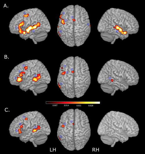
Neural activation associated with complex sentence processing in Neurotypical Adults. In an fMRI study (Europa et al., 2019), we examined brain regions activated by comprehension of syntactically complex sentences, namely passive and object cleft sentences, in 21 healthy participants, using the same task as in Barbieri et al. (2019). Results showed peak activation in the left Inferior Frontal Gyrus (IFG), Middle Frontal Gyrus (MFG), Superior Parietal Lobule (SPL), Supplementary Motor Area (SMA), temporal pole and bilateral occipital regions for all sentences over a control condition (Figure 3, top) consisting of visually scrambled pictures and auditory reversed speech. Noncanonical (compared to canonical) sentences showed peak activity in the left pars opercularis of the IFG (opIFG), the MFG, the posterior Middle Temporal Gyrus (pMTG), and occipital regions. Comparison of object cleft vs. passive sentences yielded significant activation in the left opIFG, pMTG, medial Superior Frontal Gyrus (mSFG) (Figure 3, middle) and insula (Figure 3, bottom), with no significant activation derived from the opposite contrasts.